Focal recruitment of neutrophils (red) to glomeruli (blue)
Blood vessels in the urinary bladder
Neutrophils (red) and Macrophages (green) communicate to phagocytose Debris
Spatial distribution of Neutrophils (red) and Macrophages (green) in the inflamed skin
Crosstalk of macrophages and neutrophils
Neutrophils are continuously generated within the bone marrow by myeloid precursors and their half-life is limited to a few hours. Their main function is to patrol vessels and vigilantly seeking out for indications of an incipient inflammation. Recruitment of neutrophils is induced by stimuli, such as chemokines, leukotrienes and formyl peptides. Interstitial migration seems to be facilitated by integrins, actin polymerisation, chemokine receptors, and Matrix Metalloproteinases. Once recruited from the bloodstream, neutrophils cross the dense macrophage network and acquire essential properties for optimal anti-microbial defence. Our aim in to understand neutrophil migration under inflammatory conditions.
Pathogenesis of postoperative Ileus
Localized abdominal surgery can lead to disruption of motility in the entire gastrointestinal tract (postoperative ileus). Intestinal macrophages produce mediators that paralyze myocytes, but it is unclear how the macrophages are activated, especially those in unmanipulated intestinal areas. We have shown already that intestinal surgery activates intestinal CD103(+)CD11b(+) dendritic cells (DCs) to produce interleukin-12 (IL-12). This promotes interferon-γ (IFN-γ) secretion by CCR9(+) memory T helper type 1 (T(H)1) cells which activates the macrophages. These findings indicated that postoperative ileus is a T(H)1 immune-mediated disease and identified potential targets for disease monitoring and therapy.
Macrophage-induced Fibrosis
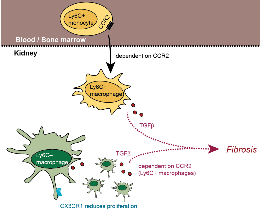
Macrophages in non-lymphoid organs are heterogeneous and specific subsets can be defined. The kidney harbors a dense network of CX3CR1+ macrophages, which directly contributes to innate immunity and might originate from blood monocytes. Nevertheless, the mechanisms that regulate migration and differentiation of these macrophages and their progenitors in homeostasis and inflammation are completely unclear. Renal macrophages depend on the chemokine receptor CCR2 and partially on CX3CR1. We are investigating these cells in the development of renal fibrosis by unilateral ureteral obstruction.
Recruited Monocytes in Infection-Induced Fibrosis
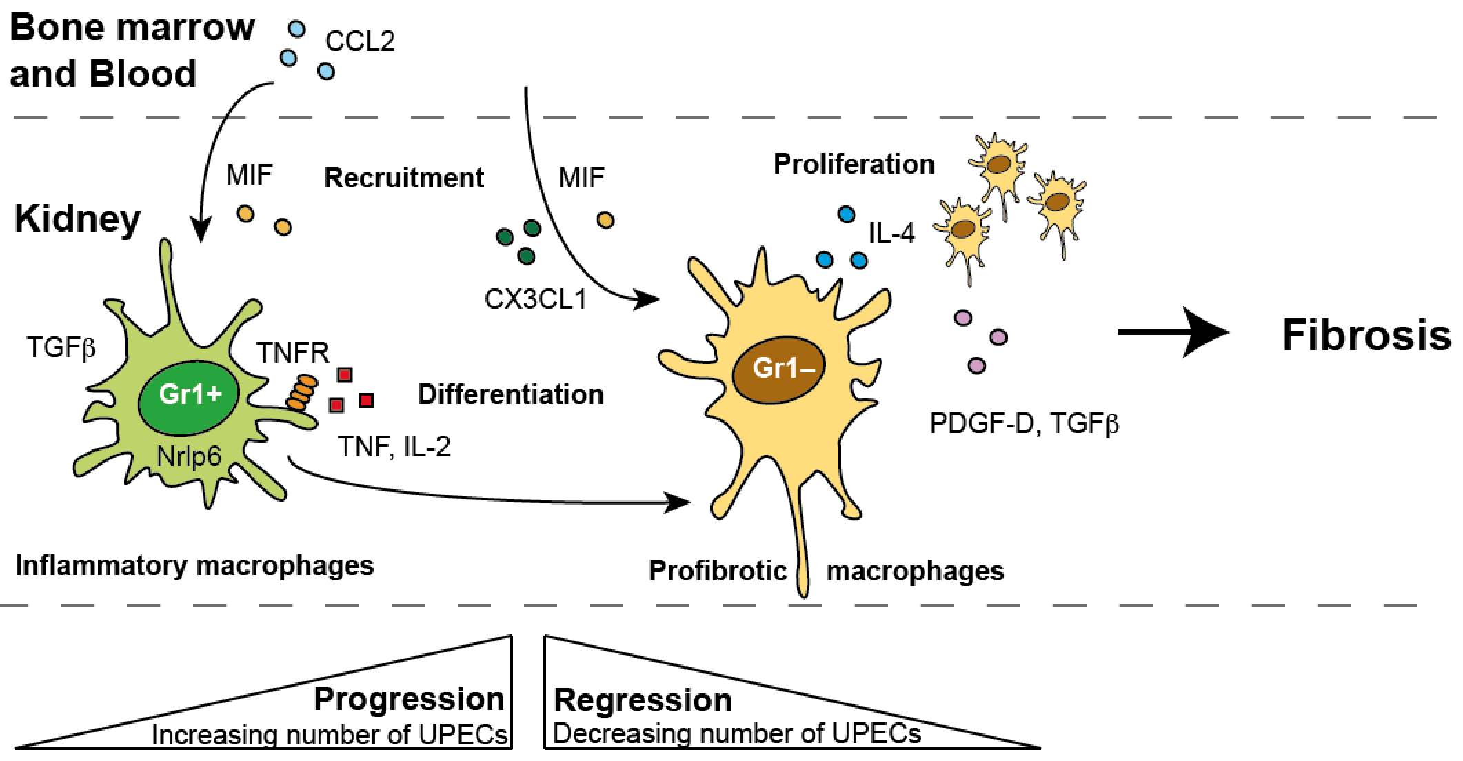
Urinary tract infections such as cystitis, urethritis and pyelonephritis are one of the most common bacterial infections worldwide. They are caused by Uropathogenic strain of E. Coli. Chronic pyelonephritis results in inflammation, acute fever, nausea and diarrhea and in long-term and inevitably renal fibrosis and furthermore kidney failure. Macrophages have been known to play an important role in fibrosis of other organs but their role in pyelonephritis- induced fibrosis is unclear. We have developed a mouse model for pyelonephritis-induced fibrosis and analyzed renal inflammation and fibrosis by flow cytometry and histology. Our major goal is to understand the molecular mechanisms in this infection and in the development of pyelonephritis-induced renal fibrosis.
Circulating Monocytes in Shiga Toxin Infection
Shiga toxin (Stx)- producing enterohemorrhagic Escherichia coli (STEC) are the primary cause of hemolytic uremic syndrome (HUS), a disease characterized by hemolytic anemia, thrombocytopenia and acute kidney failure. Endothelial cells were shown to be the main target of cytotoxic effects of Stx leading to endothelial damage, recruitment of leukocytes, and thrombus formation. However, the specific role of neutrophils and monocytes in the pathogenesis of HUS remains unclear. We are investigation the role of monocytes in this disease to unravel the monocyte-mediated pathogenesis.


